
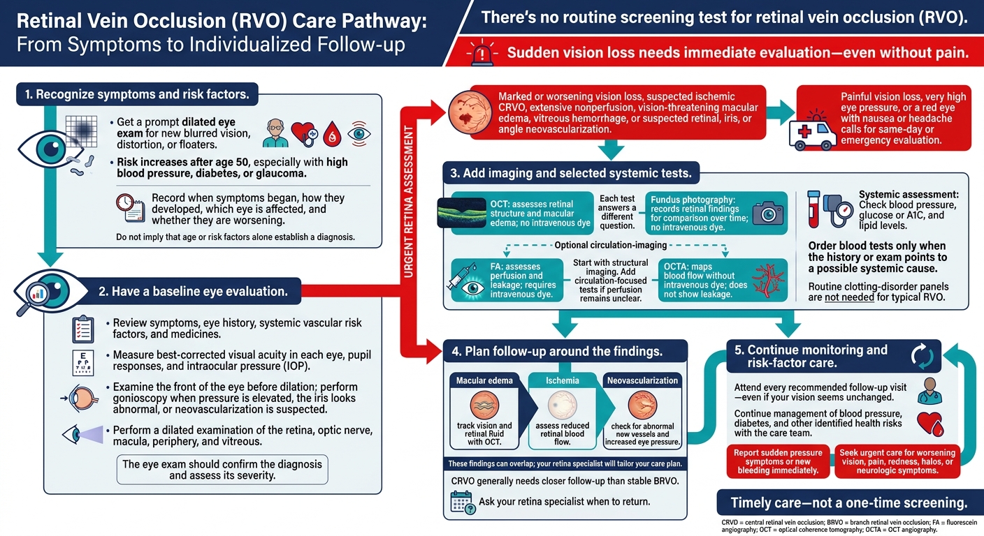
There’s no routine screening test for retinal vein occlusion (RVO). My main advice: get a prompt dilated eye exam for new blurred vision, distortion, or floaters. <u>Sudden vision loss needs immediate evaluation – even without pain.</u>
Risk increases after age 50, especially with high blood pressure, diabetes, or glaucoma. Here’s what I’ll cover:
- The first exam: How vision checks, eye pressure measurements, and a dilated exam guide diagnosis and referral.
- Imaging and blood tests: When OCT, retinal photos, circulation scans, and selected lab tests help.
- Follow-up: How swelling, reduced blood flow, and abnormal new vessels shape your visit schedule.
The goal is timely care, not a one-time test – plus control of health risks and follow-up even when vision seems unchanged.
Retinal Vein Occlusion: Evaluation and Follow-Up
Retinal Vein Occlusion
sbb-itb-929a4e2
Initial Eye Evaluation and Referral
When RVO is suspected, the eye exam should confirm the diagnosis and assess its severity.
Medical History and Baseline Eye Exam
Record when vision changes began, how they developed, whether they affect one or both eyes, and whether they are worsening. Ask about blurred or distorted central vision, blind spots, flashes, floaters, and pain. Review any history of RVO, diabetic or hypertensive eye disease, glaucoma, recent cataract or retinal surgery, eye trauma, and recent intravitreal injections. Check systemic vascular risk factors and all medicines, especially anticoagulants and antiplatelet drugs.
Check blood pressure and work with primary care to assess diabetes and lipid levels. Order blood tests only when the history or exam points to a possible systemic cause.
Measure best-corrected visual acuity in each eye, check pupil responses, and measure intraocular pressure (IOP) in both eyes. A relative afferent pupillary defect suggests severe retinal ischemia. Before dilation, use a slit-lamp exam to check for anterior chamber inflammation, iris neovascularization, and other anterior-segment findings that could mimic or complicate RVO. Perform gonioscopy if pressure is elevated, the iris looks abnormal, or neovascularization is suspected.
The dilated exam should assess the retina, optic nerve, macula, periphery, and vitreous. BRVO usually causes sectoral hemorrhage along a branch vein’s distribution. CRVO tends to cause hemorrhage across more of the retina, along with venous congestion. Look for retinal edema, poorly perfused retina, vitreous hemorrhage, and abnormal new vessels. Use imaging to document edema and nonperfusion when the exam alone is not enough. These findings guide the need for urgent retina care.
When Urgent Retina Care Is Needed
Prompt retina assessment is needed for marked or worsening vision loss, suspected ischemic CRVO, extensive nonperfusion, vision-threatening macular edema, vitreous hemorrhage, or suspected retinal, iris, or angle neovascularization. Painful vision loss, very high eye pressure, or a red eye with nausea or headache calls for same-day or emergency evaluation. Iris or angle neovascularization is especially concerning because it can block fluid drainage and increase eye pressure. Base urgency on the exam findings.
The referral should clearly document:
- Symptom onset and progression, the affected eye, best-corrected visual acuity, pupil findings, IOP, anterior-segment and gonioscopy findings, and whether dilation was completed.
- The RVO pattern, key hemorrhage or edema findings, any retina that appears ischemic, vitreous hemorrhage, and any retinal, disc, iris, or angle neovascularization.
- Available fundus photographs and OCT scans, relevant medicines, blood pressure, diabetes status, and major systemic disease.
Specify how closely the patient needs follow-up based on vision, edema, ischemia, pressure, and neovascularization. Active macular edema or high-risk ischemic findings generally call for closer review than stable BRVO without edema. Give clear instructions to seek urgent care for worsening vision, pain, redness, halos, or neurologic symptoms.
Retinal Imaging and Blood Tests
Each test answers a different question. After the dilated exam, imaging helps define retinal damage and the extent of edema and ischemia. Blood tests help identify systemic risk factors.
| Test | Intravenous dye? | Findings detected | Key limitations | Typical uses |
|---|---|---|---|---|
| OCT | No | Macular edema, intraretinal cysts, subretinal fluid, retinal thickness, and structural damage | Shows retinal structure, but not leakage or full perfusion | Baseline diagnosis, monitoring macular edema, and checking treatment response |
| Fundus photography | No | Retinal hemorrhages, venous dilation and tortuosity, cotton-wool spots, macular changes, and visible neovascularization | Flat image; media opacity, hemorrhage, or a limited field can hide findings | Setting a baseline, documenting progression, and comparing findings over time |
| Fluorescein angiography (FA) | Yes | Delayed filling, capillary nonperfusion, leakage, vessel staining, macular ischemia, and neovascularization | Requires an injection; dye reactions are possible; hemorrhage may block the view; short image-capture window; two-dimensional images | Clarifying perfusion, ischemia, unexplained vision loss, or suspected neovascularization; planning treatment |
| OCT angiography (OCTA) | No | Flow in superficial and deep capillary plexuses, capillary nonperfusion, an enlarged foveal avascular zone, and vascular abnormalities | Does not show leakage or pooling; motion, blink, segmentation, projection, and shadowing artifacts can affect interpretation; scan coverage may be limited | Dye-free assessment of macular perfusion and follow-up when dye-free imaging is preferred |
Start with structural imaging. Add circulation-focused tests if perfusion remains unclear.
OCT and Fundus Photography
Use OCT to set a baseline and track treatment response. Review the scan – not just the thickness number. Use the same device and scan pattern for follow-up, and assess findings alongside visual acuity, the fundus examination, and image quality.
Fundus photographs create a lasting record for comparison. Wide-field imaging extends that record beyond the posterior pole to the peripheral retina.
Fluorescein Angiography and OCT Angiography
Use FA when the main question involves perfusion or leakage. Choose OCTA when you need dye-free flow mapping. Wide-field FA can show peripheral nonperfusion, though dense hemorrhage may hide it until the bleeding partly clears.
OCTA maps blood flow by retinal layer. Before interpreting an area as nonperfused, check for artifacts and confirm that scan coverage is adequate. The clinical question and image quality guide whether to use FA, OCTA, or both.
Before FA, tell the team about any previous fluorescein reaction and relevant medical history, including pregnancy. The injection may cause brief nausea, temporary yellow skin discoloration, or bright-yellow urine. Serious allergic reactions are rare, but the team must be ready to provide emergency care.
Fluorescein is not the same as iodinated CT contrast. Still, discuss any previous reaction to contrast or an injection.
Blood Pressure and Selected Lab Tests
Check blood pressure, glucose or A1C, and lipid levels to guide risk-factor treatment. A complete blood count may help investigate anemia, abnormal blood-cell counts, or possible blood-viscosity problems.
Younger patients and those with bilateral, recurrent, or atypical RVO may need selected hematologic or inflammatory tests, coordinated with primary care or hematology. Routine clotting-disorder panels are not needed for typical RVO.
Planning Follow-Up Based on RVO Risk
After the baseline exam and imaging, follow-up focuses on edema, ischemia, and neovascularization. These findings can overlap, so your retina specialist will tailor your care plan to your needs.
| Monitoring category | Examination priorities | Triggers for earlier assessment |
|---|---|---|
| Stable disease | Compare visual acuity, IOP, and dilated retinal exam findings with prior visits | New symptoms |
| Macular edema | Check visual acuity and use OCT to track fluid changes | Increasing fluid or declining vision |
| Ischemic RVO | Use a dilated retinal exam and fluorescein angiography to assess nonperfusion and hemorrhages | Worsening nonperfusion or extensive new retinal hemorrhages |
| Suspected neovascularization | Examine the iris, drainage angle, retina, and optic disc, and check IOP | Abnormal vessels, vitreous hemorrhage, or a sudden increase in IOP |
Follow-Up Timing for CRVO and BRVO
Your follow-up schedule depends on ischemia, macular edema, and neovascularization. CRVO generally needs closer follow-up than stable BRVO. Ask your retina specialist when to return. At each visit, they should check whether edema, ischemia, or new blood vessel growth is worsening.
Monitoring Edema, Ischemia, and New Blood Vessels
The table above outlines what each visit should assess. Follow-up includes visual acuity testing, IOP measurement, and a dilated exam. OCT helps track macular edema, while fluorescein angiography assesses nonperfusion or neovascularization.
Report sudden pressure symptoms or new bleeding immediately. New blood vessels can bleed or contribute to glaucoma.
Conclusion: Get Prompt Care and Keep Follow-Up Visits
After diagnosis and follow-up planning, focus on timely care – not a one-time screening. RVO screening depends on symptoms and risk factors, and no single test can rule it out. A normal exam does not remove future risk or restore lost vision.
Before your visit, gather your medication list, relevant records, previous eye scans, and a symptom timeline noting when symptoms started and which eye is affected. Arrange a ride home after dilation and bring sunglasses. Blurred vision and light sensitivity can last several hours.
Denver-area patients can schedule a prompt eye evaluation and retina care with Denver Eye Surgeons. Seek immediate assessment for sudden vision loss, and attend every recommended follow-up visit – even if your vision seems unchanged.
FAQs
Can RVO develop without noticeable symptoms?
Yes. RVO and other retinal conditions can develop without noticeable symptoms, damaging the retina before you notice vision changes or discomfort. These conditions may go undetected until vision loss is severe or permanent.
Regular, comprehensive dilated eye exams are the only effective way to detect these problems early, so treatment can begin when it has the best chance of success.
Could RVO affect my other eye?
Retinal vein occlusion usually affects one eye, but both eyes need monitoring. High blood pressure and blood clotting problems can affect blood vessel health, so your eye doctor will also review your overall health during your exam.
Regular, thorough eye exams at Denver Eye Surgeons help spot changes in either eye, support care for health conditions that affect the whole body, and protect your long-term vision. Report any new symptoms or vision changes to your care team immediately.
Does RVO increase my risk of stroke?
The information provided does not explain whether retinal vein occlusion (RVO) increases your risk of stroke. If you have concerns about your overall health or stroke risk, talk with your primary care physician or a specialist.
Denver Eye Surgeons offers retina care and treatments for retinal diseases and can assess your retinal health. Call 303-279-6600 to schedule an eye exam.