
Clear vision doesn’t mean diabetes has spared your retina. My main takeaway: keep your eye exams even when you see well. A dilated exam and an OCT scan help your doctor choose between monitoring, injections, laser treatment, and surgery.
Here’s how those findings guide care:
- Early, stable damage: Scheduled exams may be enough.
- Macular swelling: Vision tests and OCT help guide observation, injections, or selected laser treatment.
- Abnormal vessels, bleeding, or retinal pulling: You may need laser, injections, or surgery.
- After treatment: Repeat exams show whether to continue or change the plan. The first check after an anti-VEGF injection is often about 4 weeks later.
I’d treat follow-up as part of treatment, not an optional check. <u>Sudden vision loss, a curtain-like shadow, or flashes with new floaters need immediate eye care.</u>
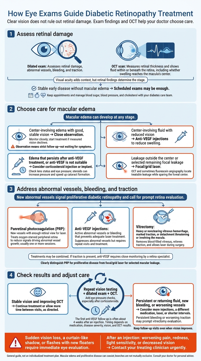
How Eye Exams Guide Diabetic Retinopathy Treatment
Step 1: Assess Retinal Damage and Plan Follow-Up
What Dilated Exams and OCT Reveal
Retinal findings guide the choice between monitoring and treatment. Your doctor examines the macula – the area responsible for sharp central vision – plus the optic nerve, retinal vessels, peripheral retina, and vitreous.
Microaneurysms are tiny bulges in blood vessels; hemorrhages are areas of bleeding. Hard exudates are lipid deposits caused by leakage, while cotton-wool spots indicate reduced blood supply. The number and location of these findings, along with swelling, venous changes, and abnormal new vessels, help determine disease severity. Visual acuity adds context, but retinal findings determine the stage.
OCT measures retinal thickness and shows fluid within or beneath the retina. It helps confirm whether swelling reaches the macula’s center. Retinal photography can document disease, but abnormal, incomplete, or ungradable images need an in-person evaluation.
How Disease Stage Guides Exam Timing
The intervals below are general planning guides. Your doctor may schedule visits sooner if retinopathy is progressing, macular edema is present, or pregnancy or prior treatment changes your risk.
| Disease stage | Key findings | Approximate monitoring interval | Next step |
|---|---|---|---|
| No retinopathy | No diabetic retinal lesions or macular edema | Usually annually; 1–2 years for selected low-risk patients with repeated normal exams and glycemic indicators at goal | Routine follow-up |
| Mild nonproliferative | Microaneurysms, with few or no other lesions | About 6–12 months | Routine ophthalmic follow-up; earlier assessment for suspected edema or progression |
| Moderate nonproliferative | More hemorrhages or microaneurysms, cotton-wool spots, hard exudates, or venous changes | About 3–6 months | Ophthalmology evaluation |
| Severe nonproliferative | Extensive hemorrhages or microaneurysms, venous beading, or intraretinal microvascular abnormalities | About 1–3 months | Prompt retina evaluation |
| Proliferative | Abnormal new vessels, vitreous or preretinal hemorrhage, or traction | Treatment-dependent; close follow-up | Prompt retina evaluation and treatment planning |
Macular edema can develop at any stage. Fluid involving the center of the macula may call for earlier assessment. Sudden floaters, flashes, a curtain or shadow, or abrupt vision loss need urgent care. Report new central blur or distortion promptly.
When Monitoring Is Enough
Stable early disease without macular edema often calls for scheduled exams rather than an eye procedure. Monitoring is active care: keep your appointments and work with your diabetes care team to manage blood sugar, blood pressure, and cholesterol.
Bring your A1C, how long you’ve had diabetes, pregnancy status, and records of any prior injections, laser treatment, or surgery. These details help your doctor decide when you should return.
If OCT shows center-involving macular edema, the next step is choosing observation, injections, or macular laser.
sbb-itb-929a4e2
Goals of Therapy When Treating Diabetic Retinopathy
Step 2: Choose Treatment for Macular Edema
When OCT shows edema involving the center of the macula, exam results help your doctor decide whether to monitor or treat it. OCT shows where fluid is and whether it persists; visual acuity testing measures its effect on vision.
Compare Observation, Injections, and Macular Laser
| Option | Findings that guide the decision | Action |
|---|---|---|
| Close observation | Center-involving edema with good, stable vision | Monitor closely; start treatment if measured vision declines. |
| Anti-VEGF injections | Center-involving fluid with reduced vision | Give injections to reduce swelling. |
| Corticosteroid injection or implant | Edema that persists after anti-VEGF treatment, or cases where anti-VEGF is not suitable | Consider steroids after checking lens status and eye pressure. |
| Focal/grid laser | Leakage outside the center or selected areas of remaining focal leakage | Use OCT and sometimes fluorescein angiography to locate treatable leakage while sparing the foveal center. |
Once treatment begins, repeat vision tests and OCT scans show whether swelling is improving or the plan needs to change.
When vision is good, observation can work as well as early treatment. But observation means strict follow-up – not waiting for symptoms. Treatment should begin if testing shows that vision has declined.
Anti-VEGF medicines include aflibercept, ranibizumab, and bevacizumab. Treatment usually takes several injections, with vision tests and OCT guiding further doses.
Exam results also help your doctor decide whether remaining leakage needs laser treatment or persistent edema needs steroids. Before steroid treatment, your doctor checks lens status and eye pressure because steroids can increase pressure and speed up cataract formation.
Focal/grid laser may supplement injections. It is different from panretinal laser, which targets areas of poor blood supply in the peripheral retina in proliferative disease – not macular edema.
Step 3: Address Abnormal Vessels, Bleeding, and Traction
When an exam shows new vessels, bleeding, or traction, treatment shifts from controlling macular swelling to preventing repeat bleeding and retinal detachment. The findings help guide the choice of laser, injections, or surgery.
New abnormal blood vessels on the retina or optic disc signal proliferative diabetic retinopathy (PDR). These findings call for prompt evaluation by a retina specialist – before major vision loss occurs.
Compare Panretinal Laser, Anti-VEGF, and Vitrectomy
Wide-field imaging and, when needed, fluorescein angiography map areas with poor blood flow and help determine whether laser treatment is possible. If bleeding blocks the view, B-scan ultrasound checks whether the retina is attached and helps assess the need for surgery.
| Treatment | Retinal problem | Exam findings supporting it | Why it matters |
|---|---|---|---|
| Panretinal photocoagulation (PRP) | Proliferative disease | Visible new vessels on the retina or optic disc, with enough retinal view to apply laser | Treats oxygen-starved retina to reduce the signals that drive abnormal vessel growth. |
| Anti-VEGF injections | Active abnormal vessels, sometimes with macular edema | New vessel growth or bleeding that prevents adequate laser treatment | Suppresses abnormal vessels but requires repeat visits and treatment. |
| Vitrectomy | Heavy or nonclearing vitreous hemorrhage; marked traction or detachment | Blood blocks the view of the retina, or detachment threatens or involves the macula | Removes blood-filled vitreous, relieves traction, and allows laser treatment during surgery. |
PRP usually takes one or more sessions to reduce the retinal signals that drive abnormal vessel growth.
These treatments often work together rather than alone. Injections may quiet active vessels before PRP, and vitrectomy may include laser treatment. If traction is present, anti-VEGF requires close monitoring by a retina specialist.
Step 4: Check Treatment Results
How Follow-Up Exams Guide Treatment Changes
After injections, laser treatment, or surgery, follow-up exams help your clinician see how your retina is responding. They compare visual acuity, a dilated retinal exam, and repeat OCT scans with earlier results. These checks track vision, retinal thickness, fluid, bleeding, and abnormal blood vessels. Eye-pressure checks are especially important after corticosteroid injections or implants because steroids can increase intraocular pressure.
These results help guide whether to continue or change treatment. The first anti-VEGF follow-up is often about 4 weeks after an injection. Timing depends on the medication, disease severity, vision, and OCT results. If vision stays stable and OCT results improve, your clinician may continue treatment or allow more time between visits. Keep follow-up visits even when vision improves. Changes on OCT scans or eye exams can appear before symptoms return.
Retinal fluid that persists or returns may call for more injections, a different medication, laser treatment, or shorter treatment intervals. New bleeding or worsening abnormal vessels may also need additional treatment. Vitreous bleeding that persists or doesn’t clear, scar tissue, or worsening traction may prompt an evaluation for vitrectomy. Your clinician bases these decisions on changes across exams – not a single scan or vision score.
When Vision Changes Need Urgent Care
Follow-up visits also check for warning signs that need urgent attention. After treatment, report new or increasing floaters or blurred vision promptly. Sudden vision loss, a curtain-like shadow, or flashes with new floaters require immediate eye evaluation. After an injection, contact your treating clinician urgently if you develop worsening pain, redness, light sensitivity, or decreased vision. Don’t wait for your next appointment.
Before leaving each visit, confirm your next appointment and treatment plan. Ask which symptoms call for an earlier check and how to reach the care team urgently.
Conclusion: Let Exam Findings Guide Your Care
Retinal findings and OCT help your doctor decide whether to monitor diabetic retinopathy or treat it with injections, laser, or surgery. Follow-up exams show whether treatment is working.
Before your visit, gather your medication list, recent A1C result, blood pressure history, and records of past eye treatments. These details help your doctor put the exam findings in context. Ask which retinal finding is guiding treatment and what change your doctor expects to see.
Denver Eye Surgeons in Denver offers diabetic eye care and retina services. Share your records so the team can compare today’s findings with your treatment history.
FAQs
Why might my OCT improve but my vision stay blurry?
Your optical coherence tomography (OCT) scan may show less retinal swelling or fluid, but your retina may still need time to heal and regain function.
Other eye conditions, such as cataracts, can keep your vision blurry even as your retina improves. Blood sugar fluctuations can also temporarily change the shape of your eye’s lens, causing blurred vision even when your retina is stabilizing.
How does my doctor decide when to switch treatments?
Your eye doctor checks how well your current treatment is working, including whether fluid buildup remains despite anti-VEGF injections. If treatment isn’t delivering the expected results, they may switch medications or add laser therapy.
At Denver Eye Surgeons, these decisions depend on regular exams, your retinal health, your overall medical condition, and how you’ve responded to past treatments. Your care stays tailored to you as your needs change.
Can I stop injections once my vision improves?
As your vision improves and your condition stabilizes, you may need anti-VEGF injections less often. But diabetic retinopathy still needs long-term care to help prevent it from returning.
Keep following your Denver Eye Surgeons doctor’s treatment plan, even if you’re seeing better. Regular follow-up exams let your doctor check how well treatment is working and decide whether it’s safe to adjust your injection schedule or continue maintenance therapy to prot