
Adaptive optics (AO) retinal imaging corrects distortions in the eye to show tiny retinal structures – sometimes about 2 micrometers across. My main takeaway: it adds cellular detail, but it does not replace a dilated eye exam or standard OCT.
Here’s what I’d keep in mind:
- What it shows: Individual light-sensing cells and, with some systems, tiny blood vessels and other retinal structures.
- Why it’s used: Mainly to study retinal disease and track cellular changes during research and treatment trials.
- Where it falls short: Eye movement, cloudy lenses, and differences between devices can affect results. Sharper images alone don’t establish a diagnosis.
- What to ask before booking: Will the scan affect my care? Is dilation needed? What will I pay, and does insurance cover it?
<u>I’d confirm availability and costs with the imaging center first.</u> AO is mainly offered at research centers and select specialty practices; Denver Eye Surgeons can confirm whether it offers the scan.
Adaptive Optics for Retinal Imaging by Scot S. Olivier
sbb-itb-929a4e2
How Adaptive Optics Retinal Imaging Works
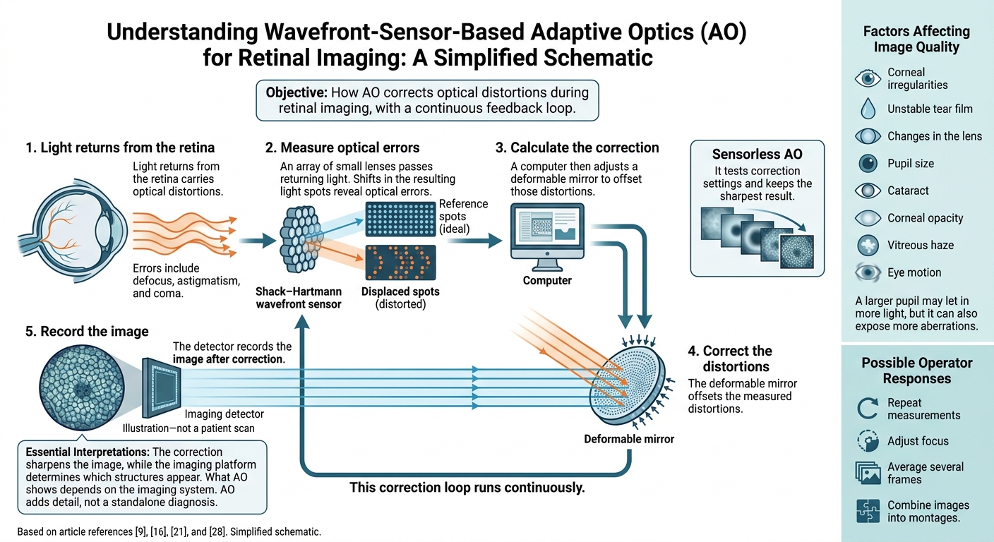
How Adaptive Optics Retinal Imaging Works
Measuring and Correcting Optical Imperfections
A Shack–Hartmann wavefront sensor passes light returning from the retina through an array of small lenses. Shifts in the resulting light spots reveal optical errors, such as defocus, astigmatism, and coma. A computer then adjusts a deformable mirror to offset those distortions before the detector records the image. This correction loop runs continuously. Sensorless AO takes a different approach: it tests correction settings and keeps the sharpest result. The correction sharpens the image, while the imaging platform determines which structures appear.
Corneal irregularities, an unstable tear film, and changes in the lens can blur light. Pupil size also affects fine detail. A larger pupil may let in more light, but it can also expose more aberrations. Cataract, corneal opacity, vitreous haze, and eye motion can reduce image quality. To address these issues, operators may repeat measurements, adjust focus, average several frames, or combine images into montages.
Retinal Cells and Structures It Can Show
What AO shows depends on the imaging system, not simply whether it uses AO. Flood-illumination ophthalmoscopy produces a two-dimensional, face-on view of the retina. AO scanning laser ophthalmoscopy (AO-SLO) scans a focused laser spot across the retina, while confocal detection rejects out-of-focus light. AO-OCT pairs improved transverse resolution with depth-resolved, cross-sectional imaging.
Depending on the system and wavelength, images can show cone and rod photoreceptors, retinal pigment epithelial cells, capillaries, and retinal nerve fiber bundles. Rods are often harder to image because they are smaller and may return weaker signals. Some approaches can show ganglion-cell bodies, though not in every patient or with every system. Some research AO-OCT systems have reached approximately 2–3 micrometers of transverse resolution, but performance varies by instrument and eye.
How Adaptive Optics Compares With Fundus Photography and OCT
AO improves compatible imaging systems rather than replacing them. Each method helps answer a different question: where a retinal change lies, which layers it affects, or what fine cellular patterns look like. AO adds detail, not a standalone diagnosis.
| Imaging method | Primary view | Retinal detail | Typical clinical or research role | Major limitation |
|---|---|---|---|---|
| Fundus photography | Broad, face-on (en face) color or monochromatic view of the retina and optic nerve | Lesion- and vessel-level detail; usually does not show individual cells | Documentation, screening, and monitoring visible retinal changes | Limited microscopic and depth information |
| Standard OCT | Cross-sectional and volumetric view of retinal layers | Micrometer-scale layer structure and thickness | Diagnosing and monitoring macular disease, edema, and structural change | May not resolve individual photoreceptors or fine capillary architecture |
| AO ophthalmoscopy or AO-SLO | High-resolution en face view | Cellular mosaics, fine vessels, nerve fiber bundles, and selected retinal layers | Research, phenotyping, and assessment of cellular change | Limited field of view, sensitivity to fixation and ocular aberrations, and limited routine availability |
| AO-OCT | Depth-resolved, cellular-scale OCT view | High transverse resolution combined with cross-sectional structure | Research and emerging clinical applications | Technically complex, slower, and not a replacement for standard OCT |
Uses and Limits in Retinal Disease Assessment
Studying Retinal Disease and Treatment Effects
AO is mainly a research tool for studying retinal changes linked to specific diseases. Its cellular detail helps researchers track disease progression and response to treatment.
In AMD, AO shows drusen, pigment changes, photoreceptor loss, and the borders of geographic atrophy. In diabetic retinopathy, it can reveal reduced cone density, capillary changes, and microaneurysms. Cone loss increases as retinopathy advances. For inherited retinal diseases, including Stargardt disease and retinitis pigmentosa, AO maps cone and rod loss. Glaucoma researchers use it to examine nerve fiber bundles and, with some systems, ganglion-cell structures.
Clinical trials use AO to revisit the same retinal sites and measure changes in photoreceptor spacing, cone density, capillaries, or lesion shape. These measures remain experimental unless validated for a specific disease and treatment. Preserved retinal structure alone does not prove better vision or quality of life.
Advantages and Practical Limits
AO’s limits depend on the imaging system, software, operator experience, and patient characteristics.
| Advantages | Limitations |
|---|---|
| Repeated cellular imaging to track structural changes | Small sampling areas need precise alignment. Unstable fixation and fatigue can reduce reliability, and image acquisition and analysis take time. |
| Fine photoreceptor and vascular measurements | Ocular opacities, tear-film problems, and uncorrected refractive error can reduce image quality. |
| Quantitative measurements over time | Results vary across devices, operators, patients, and retinal locations. |
| Cellular measurements for clinical trials | Protocols, reference values, and analysis methods are not yet fully standardized. |
For repeat measurements, researchers should keep retinal locations, acquisition settings, and analysis methods consistent. Sampling a slightly different spot can look like biological change. Measurements from different devices should not automatically be treated as interchangeable.
Why Images Alone Cannot Establish a Diagnosis
AO samples only a small retinal area. Its findings must be considered alongside symptoms, medical history, visual acuity, a dilated exam, OCT, fundus photography, angiography when indicated, and visual fields.
An eye-care professional should interpret AO findings in the context of the full exam. Microscopic abnormalities may not point to a specific disease. More detail alone does not prove earlier diagnosis, predict vision loss, or show that a treatment helps. AO therefore serves as part of a broader retinal workup – not a replacement for standard testing.
What Patients Can Expect From Retinal Imaging
Before, During, and After Imaging
If AO imaging is part of your retinal workup, the visit is usually straightforward. Bring your eye history, symptoms, medication list, and recent test results. Follow the center’s instructions about contact lenses, makeup, and medications. Don’t stop prescribed medicines on your own. Ask whether your eyes will be dilated, bring sunglasses, and arrange a ride home if driving may be unsafe.
During the scan, you’ll usually rest your chin and forehead on supports and look at a target while the technician takes images of the target area. Staff may adjust the focus or repeat scans if you blink or move your eyes. Visit length and dilation needs vary.
Images often need processing, so results may not be ready right away. Ask when to expect them and who will review them.
Availability, Costs, and Insurance
Confirm the scan’s purpose and cost before scheduling. AO imaging is mainly available at research centers and select specialty practices. It isn’t routinely offered – or needed – for every retinal condition. Ask whether the scan is for care or research and whether it will affect your treatment.
Request the procedure name, billing code, and a written estimate. If the scan is part of research, ask whether a sponsor covers imaging costs. Also check for separate facility or image interpretation fees.
Contact your insurer to confirm coverage, prior authorization requirements, your deductible, copayments, and coinsurance. Don’t assume the scan is covered.
Retinal Care at Denver Eye Surgeons
Denver Eye Surgeons in Lakewood, Colorado, provides retina and diabetic eye care. Contact the practice directly to ask whether AO imaging is available and appropriate for your case. The key question is whether it will add useful detail to your retinal evaluation.
Conclusion: The Role of Adaptive Optics in Retinal Care
Adaptive optics (AO) provides a noninvasive view of tiny retinal structures, including photoreceptors and capillaries. It does not replace standard retinal care. Its main uses are detailed analysis, research, clinical trials, and tracking changes in selected patients – not routine diagnosis.
Reliable follow-up comparisons depend on imaging the same retinal location with the same method. Differences in alignment, image quality, or analysis can distort results.
More detail does not equal a diagnosis: specialists must interpret AO findings in the patient’s clinical context. Patients should continue recommended dilated exams and standard imaging, including OCT. AO adds microscopic detail, while standard examinations remain the basis of retinal care.
FAQs
Is adaptive optics imaging safe for my eyes?
Yes, adaptive optics retinal imaging is considered safe. It provides detailed views of the structures inside your eye without surgery or instruments entering the eye. Like optical coherence tomography (OCT), it’s designed to image these structures without harming your vision.
If you have questions about the imaging equipment used during your visit, our team at Denver Eye Surgeons can explain how it works and help you feel comfortable and informed.
When would an AO scan change my treatment?
An adaptive optics (AO) scan may change your treatment plan by showing small retinal changes in detailed images. These findings can help your doctor refine your diagnosis and decide whether you need injections, laser surgery, or another surgical procedure to stabilize or improve your vision.
At Denver Eye Surgeons, advanced imaging helps guide care tailored to your needs, including treatment adjustments over time to protect your eye health.
Can AO explain blurry vision despite a normal OCT?
Yes. Adaptive optics (AO) retinal imaging may help explain blurry vision even when an optical coherence tomography (OCT) scan looks normal. OCT shows cross-sectional views of the retina’s layers. AO offers higher-resolution views of individual cells, including photoreceptors, which can reveal subtle structural changes that standard imaging may miss.
Denver Eye Surgeons can discuss advanced diagnostic options based on your needs.